Microglial mitochondrial stress may help explain brain aging
Microglial mitochondrial stress may help explain how brain immune cells age, but the new Nature study stops well short of an Alzheimer's treatment.
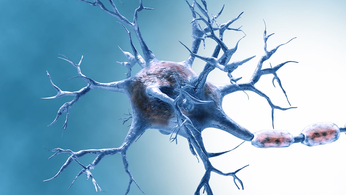
A new 2026 Nature Neuroscience study by Maria Jose Perez J and Michela Deleidi suggests that when microglia, the brain’s resident immune cells, stay locked in a mitochondrial stress program, they do more than look inflamed. In human cell models, they lost key metabolites, remodelled their lipids and slipped into a senescence-like state that changed how they communicated with neurons.
That does not mean researchers have found a treatment for Alzheimer’s disease, or even proved that this pathway is a direct driver of dementia in people. It does sharpen a mechanistic question that has been hanging over brain-aging research for years: when microglia age badly, are they simply reacting to damage around them, or can their own metabolic stress help push the rest of the brain off balance?
Here, the answer is cautious but important. The work points to the mitochondrial unfolded protein response, often shortened to UPRmt, as one route by which stressed microglia may stop doing their maintenance work and start behaving like old, dysfunctional cells. That is a more specific claim than the familiar line that neuroinflammation is involved in Alzheimer’s. It narrows the field from a vague inflammatory haze to a defined cell state with identifiable metabolic weak points.
What the new Nature paper actually showed
Its main strength is that it did not stop at one dish of isolated cells. Perez J and Deleidi first induced mitochondrial proteotoxic stress in human iPSC-derived microglia, then followed the consequences through more complex systems, including tricultures and brain organoids, to see whether the same stress program disrupted how glia and neurons interact.

What emerged was not just a generic inflammatory flare. The stressed microglia showed depletion of S-adenosyl methionine, or SAM, a central methyl donor used across cellular metabolism, along with lipid remodelling and a senescence-like signature. That combination matters because it suggests the cells were being pushed into a durable state change, not merely having a short, noisy response to injury.
On the neuron side, the findings become more than a cell-biology curiosity. In the authors’ co-culture systems, the microglial stress response was tied to disrupted neuronal-glial communication, which is precisely the kind of systems-level failure that brain-aging researchers worry about. Microglia are supposed to clear debris, prune synapses selectively and help maintain the tissue environment. If their stress program scrambles those jobs, the downstream problem is not only inflammation. It is lost support.
Those human-derived models do not solve every translational problem, but they do answer a familiar objection. Too much of the Alzheimer’s pipeline has moved from elegant mouse findings to disappointing human results. By showing the same broad stress signature in human microglia and in more tissue-like systems, the paper clears a higher bar than a single rodent experiment would have.
Why the metabolic shift matters
The finding also lands neatly inside a line of evidence that has been building for several years. A 2022 Nature Neuroscience study led by Thomas Blank linked aging microglia in mice to microbiota-driven oxidative stress and mitochondrial damage, while a 2025 review in Frontiers in Aging argued that dysfunctional mitochondria and neuroinflammation can reinforce one another across several neurodegenerative diseases.
Seen against that backdrop, the new paper feels more consequential than another entry in the crowded Alzheimer’s mechanism literature. Many brain-aging papers still describe inflammation as though it were an atmospheric condition, everywhere and nowhere at once. This one gets closer to upstream biology. If microglia under mitochondrial stress deplete SAM and rework their lipid handling, the relevant question becomes whether those metabolic changes are helping to create the senescent phenotype, or merely tagging along with it.
One reason the SAM finding stands out is that it places the problem upstream of the usual inflammatory readouts. A cell that is short on methyl donors is not just irritated. It may be failing at basic housekeeping, gene regulation and membrane maintenance. The lipid remodelling signal points in the same direction. Before a microglial cell starts broadcasting inflammatory distress, it may already have become metabolically bad at being a microglial cell.
For drug development, that distinction matters. Anti-inflammatory strategies have often looked cleaner on paper than in patients because inflammation is the final common language of many damaged tissues. A metabolic choke point is different. It gives researchers something narrower to test, even if the testing still has to happen far upstream of any clinical claim. In that sense, the study reads less like a breakthrough and more like a map annotation: look here, this part of the pathway may be load-bearing.
How close this gets to Alzheimer’s biology
Not as close as some headlines will imply, but closer than a mouse-only story. The most relevant comparison is a 2024 Nature Neuroscience paper by Noa Rachmian and colleagues, which found senescent, TREM2-expressing microglia across aging, amyloidosis and tauopathy in mouse models. In that study, senolytic treatment reduced senescent microglia, lowered inflammatory signalling and improved cognition in mice. The implication was provocative: senescent microglia might be contributing to disease, not just reflecting it.
The 2026 paper does not establish that same causal chain in humans. Instead, it does something narrower and arguably more useful at this stage. It shows that a specific mitochondrial stress response can push human microglia toward a senescence-like program and break down neuron-glia communication in human-derived experimental systems. That makes the Alzheimer’s link more plausible, but it does not yet make it actionable.
Another gain is conceptual. Alzheimer’s risk biology keeps circling back to microglia, immune signalling and tissue maintenance, not only to amyloid plaques viewed in isolation. A mechanism paper like this helps explain why. If microglia enter a bad metabolic state early enough, they may stop being reliable housekeepers long before overt neurodegeneration becomes visible. That is the sort of shift that could help connect aging, inflammation and vulnerability to later disease.
The same framing fits where the broader field has been moving. A June 2026 New Scientist report on a large Alzheimer’s genetics study noted that several newly identified risk signals again pointed toward microglial genes, reinforcing the idea that immune-cell maintenance is central, not incidental, to neurodegeneration.
Still, plausibility is not proof of risk. Perez J and Deleidi do not show that people with early Alzheimer’s, mild cognitive impairment or genetically elevated risk already carry this exact UPRmt signature in vivo. Their paper shows that the pathway is biologically credible and experimentally reproducible. That is valuable, but it belongs in the category of target validation, not clinical guidance.
What the study cannot yet tell us
For all of its sophistication, the paper still lives in the space between elegant model systems and messy human brains. iPSC-derived microglia are useful, organoids are useful, and tricultures are useful. None of them fully captures decades of human aging, vascular change, peripheral immune input or the layered pathology seen in real Alzheimer’s disease.
A second caveat is the familiar trap in senescence research: a cell can look senescent by several markers without behaving exactly like the senescent cells found in aged human tissue. That does not invalidate the work, but it should slow down any rush to translate these findings into consumer brain-health advice or near-term therapy talk. Vitalspell readers should treat this as a mechanism paper first, a disease paper second and a treatment paper not at all.
Researchers will now have to ask harder questions. Can the same metabolic signature be detected in patient tissue, cerebrospinal fluid or imaging-linked biomarkers? Does blocking this stress response preserve neuronal support, or does it merely change a laboratory phenotype? And if senescent microglia are part of the Alzheimer’s story, when in the disease course do they matter most?
The bottom line is narrower than a treatment headline. The study suggests that mitochondrial stress in microglia may be one way the aging brain starts to lose immune balance and neuronal support. It also gives researchers a tighter hypothesis to test against human tissue, biomarkers and, eventually, interventions. For now, the value is explanatory. It tells the field a little more clearly where to look when brain immune cells start acting old before the rest of the brain is ready.
References
- Perez J MJ, Deleidi M. The mitochondrial unfolded protein response in human microglia disrupts neuronal-glial communication and promotes senescence. Nature Neuroscience. 2026. https://www.nature.com/articles/s41593-026-02320-1
- Rachmian N, et al. Identification of senescent, TREM2-expressing microglia in aging and Alzheimer’s disease model mouse brain. Nature Neuroscience. 17(9). 2024. https://www.nature.com/articles/s41593-024-01620-8
- Blank T, et al. Gut microbiota drives age-related oxidative stress and mitochondrial damage in microglia via the metabolite N6-carboxymethyllysine. Nature Neuroscience. 25(5). 2022. https://www.nature.com/articles/s41593-022-01027-3
- Kopeikina KJ, et al. Dysfunctional mitochondria and neuroinflammation in neurodegenerative diseases associated with aging. Frontiers in Aging. 2025. https://www.frontiersin.org/journals/aging/articles/10.3389/fragi.2025.1615764/full
General assignment health reporter covering nutrition science, wellness trends, and clinical research. Reports from Toronto.
The Vitalspell brief
Evidence-based supplement science — weekly in your inbox.
Subscribe