How karyoptosis may help explain neuron loss in Alzheimer's
Karyoptosis in Alzheimer's may explain how proteotoxic stress turns toxic proteins into neuron loss in Alzheimer's and frontotemporal dementia.
A longstanding Alzheimer’s question has been hiding in plain sight. Researchers have spent decades mapping toxic proteins, inflamed tissue and failing cellular housekeeping systems, yet the exact step where a stressed neuron crosses into irreversible death has been harder to pin down. A 2026 Nature Communications paper by Rebecca Casterton and colleagues argues that one missing link may be karyoptosis, a form of cell death driven by nuclear breakdown after proteotoxic stress.
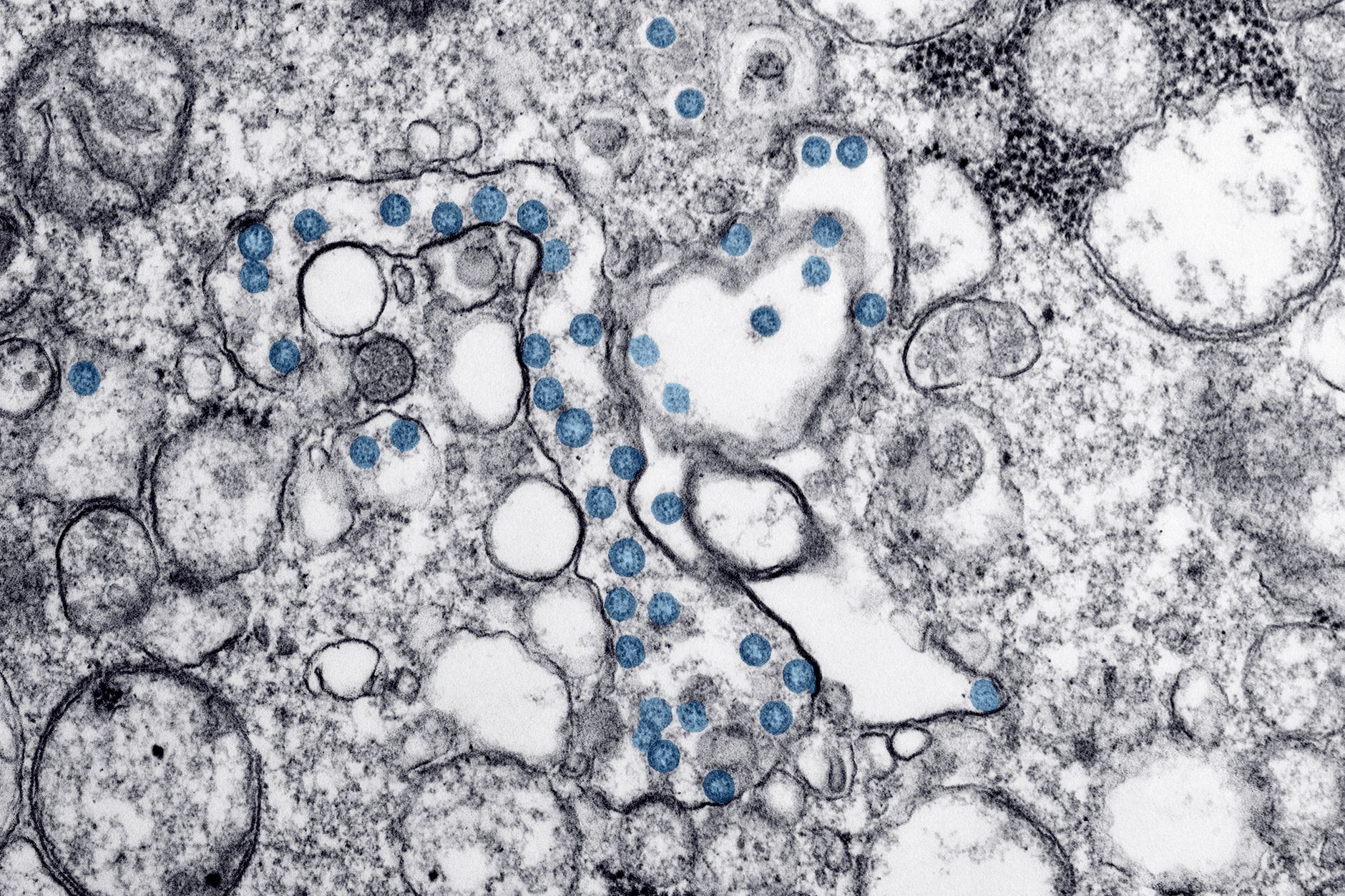
The insider view is what makes the paper interesting. Rather than treating amyloid, tau or frontotemporal-dementia aggregates as the whole story, the authors ask what those insults are doing to the cell’s final decision-making machinery. In their post-mortem frontal-cortex analysis, the team reported karyoptotic features in roughly 35 percent of cells from Alzheimer’s brains, compared with about 15 percent in healthy older-adult tissue. They also identified similar signatures in frontotemporal dementia. If that pattern holds up, dementia research gets something it badly needs: not another vague “damage pathway” but a specific route from protein stress to neuron loss.
A skeptic will read the same result more carefully. Neurodegeneration is full of cell-death labels that look clean in model systems and blur in human tissue. The images are striking, but the harder question is whether karyoptosis is truly distinct from apoptosis, necrosis or late-stage lysosomal collapse. Another question is whether the human-brain signal captures a driver of disease rather than molecular wreckage left behind after neurons are already doomed.
What the Nature Communications paper found
Casterton’s team built the case across cultured cells, rat neurons, ALS-frontotemporal-dementia models and post-mortem human tissue. The central claim is mechanistic: proteotoxic stress activates p38 MAP kinase, p38 destabilizes the nuclear-lamina protein LaminB1, and that loss of lamina integrity pushes neurons toward karyoptosis. Put simply, the paper is not just saying stressed neurons look abnormal. It is saying a specific kinase-lamina axis may organize the death program itself.

Alzheimer’s has lacked a satisfying bridge between protein accumulation and cell disappearance. Casterton and colleagues report that karyoptosis becomes visible through nuclear degeneration and expulsion of nuclear material, not just the caspase-heavy features usually associated with apoptosis. The paper’s scale is still modest by clinical standards. Still, more than 3,000 brain cells across 28 people is enough to make the observation hard to dismiss as a one-off staining artefact.
Casterton described the finding this way in a study summary released alongside the paper:
“Our study uncovers a new series of chemical events which can coordinate cell death in brain cells.”
Rebecca Casterton, King’s College London study summary
More persuasive than the dramatic label is the paper’s attempt to connect the pathway to an intervention point. The authors argue that interfering with the p38-LaminB1 interaction slowed the march toward cell death in experimental systems. That is still a long way from a drug, and farther still from a human treatment. Even so, it partly answers the insider perspective’s first question. The paper does more than classify a corpse. It suggests a lever, at least in early-stage models, that may preserve stressed neurons before the damage becomes terminal.
Why the skepticism matters
Cell-death taxonomy is a graveyard of overconfident first drafts. What looks distinct in a dish can collapse into overlap once researchers compare multiple tissues, disease stages and stress pathways. Skepticism here is not academic throat-clearing. It is the load-bearing challenge to the whole argument: does karyoptosis genuinely separate from other death programs, or is it a vivid endpoint inside a broader degenerative continuum?

There are reasons to take the category seriously. Its emphasis on LaminB1 instability, p38 signalling and nuclear-material expulsion gives the pathway a shape that is more specific than a generic “dying cell” description. Post-mortem tissue, however, is a hard place to prove causality. A frontal-cortex snapshot cannot reveal whether karyoptosis is an early, disease-driving event or a late event that appears after proteostasis, inflammation and lysosomal failure have already done most of the destructive work.
Timing matters especially in Alzheimer’s. A mechanism that shows up only after large numbers of neurons are already in crisis can still be biologically real, but it is less useful as a biomarker and less attractive as a therapeutic target. The skeptic’s demand for independent cohorts, earlier-stage tissue and cleaner separation from apoptosis is not obstruction. It is the minimum price of admission for any claim that this pathway helps explain why neurons die, rather than how dead neurons look once the disease has run for years.
Where lysosome repair enters the picture
From the analyst perspective, the paper matters because it fits a broader literature on proteostasis failure. In a 2025 Nature Cell Biology study, Chou and colleagues showed that transdifferentiated neurons from people with Alzheimer’s accumulate proteotoxic deposits, suffer constitutive lysosomal damage and struggle with ESCRT-mediated lysosomal repair. That study did not name karyoptosis. It did, however, make a strong case that neurons in ageing and Alzheimer’s can live in a state of chronic failed cleanup.
Read next to the new paper, a plausible sequence comes into view. Protein aggregates accumulate. Lysosomes and related repair machinery start to fail. Stress signalling rises. The nuclear lamina becomes unstable. Then neurons tip into a terminal program that looks like karyoptosis. That chain is not yet proven end to end, and Vitalspell readers should resist the temptation to treat it as settled fact. It is still a better hypothesis than the field’s older habit of gesturing at “toxic proteins” as if the middle steps were self-explanatory.
A second clue comes from a 2026 Nature paper on the LASER complex and lysosome repair. Claire Goul and colleagues describe how damaged lysosomes recruit ESCRT machinery through a newly defined damage-sensing assembly. That is not an Alzheimer’s paper, but it sharpens the background question raised by Casterton’s work. If cells have a repair apparatus for damaged lysosomes, one reading of karyoptosis is that it marks what happens when repair systems no longer keep up.
This interpretation only partially answers the analyst’s main question about whether karyoptosis sits downstream of lysosomal-repair failure. The evidence is convergent, not definitive. Yet convergence matters in neurodegeneration, where no single paper usually solves the biology on its own. Casterton’s study adds a candidate end-stage mechanism. Chou’s paper helps explain why stressed neurons might be pushed toward it. The lysosome-repair work supplies cellular infrastructure that makes the story coherent instead of merely speculative.
What this changes, and what it does not
The immediate change is conceptual clarity. The karyoptosis paper gives dementia researchers a more testable description of neuron death than the catch-all language that often surrounds Alzheimer’s pathology. That alone is useful. A mechanism can be wrong in part and still move the field forward if it forces cleaner experiments, better biomarkers and sharper questions about timing. For patients or families, though, the right reading is restraint. This is not a treatment breakthrough. It is a map revision.
Fanto made that restraint explicit in the same study summary, while still sketching the long-term hope:
“By specifically targeting the interaction between p38 MAP kinase and LaminB1 we may slow down the process of cell death, buying time for more pinpointed therapies.”
Manolis Fanto, King’s College London study summary
Here the important words are “may” and “buying time.” They mark the distance between a mechanistic foothold and a usable therapy. The next tests are easy to state and hard to do: replicate the human-tissue signal, show that the pathway appears early enough to matter, and demonstrate that modulating it preserves neurons without breaking essential stress responses elsewhere in the brain. If those tests fail, karyoptosis becomes a provocative side road. If they succeed, Alzheimer’s research may finally have a clearer answer to one of its oldest questions.
For now, the paper’s strongest contribution is that it makes neuron loss in dementia look less mystical. Toxic proteins are still part of the story, but not the whole plot. The harder problem has been explaining how a stressed neuron actually dies. Karyoptosis may not be the final answer. It may, however, be the first recent answer that connects proteotoxic stress, nuclear breakdown and brain-cell loss in a way the field can genuinely interrogate.
References
- Casterton R, Martinez-Cotrina A, Barnard J, et al. Karyoptosis mediates cell death and neurodegeneration upon proteotoxic stress. Nature Communications. 2026. https://doi.org/10.1038/s41467-026-73802-w
- Chou CC, Vest R, Prado MA, et al. Proteostasis and lysosomal repair deficits in transdifferentiated neurons of Alzheimer’s disease. Nature Cell Biology. 2025. https://www.nature.com/articles/s41556-025-01623-y
- Goul CS, et al. LASER couples damage sensing to ESCRT assembly for lysosome repair. Nature. 2026. https://www.nature.com/articles/s41586-026-10604-6
General assignment health reporter covering nutrition science, wellness trends, and clinical research. Reports from Toronto.
The Vitalspell brief
Evidence-based supplement science — weekly in your inbox.
Subscribe